Leading expert in cerebrovascular and skull base neurosurgery, Dr. Philip Theodosopoulos, MD, explains the critical role of continuous anatomical study in achieving surgical excellence. He details his rigorous research methods using CT scans and cadaveric dissections to master complex skull base anatomy. Dr. Philip Theodosopoulos, MD, emphasizes that normal anatomy can challenge even the most experienced surgeons. He advocates for a return to foundational anatomical principles to push the boundaries of surgical skill and ensure patient safety.
More from MD Education
More from Diagnostic Detectives Network

Mastering Surgical Anatomy for Superior Skull Base Neurosurgery
Jump To Section
- Anatomy: The Foundation of Surgical Excellence
- Research Methods in Anatomical Study
- Why Normal Anatomy is a Surgical Challenge
- Cadaveric Dissection for Skill Development
- Endoscopic Approaches and Anatomical Perspectives
- The Imperative of Continuous Learning for Surgical Mastery
- Full Transcript
Anatomy: The Foundation of Surgical Excellence
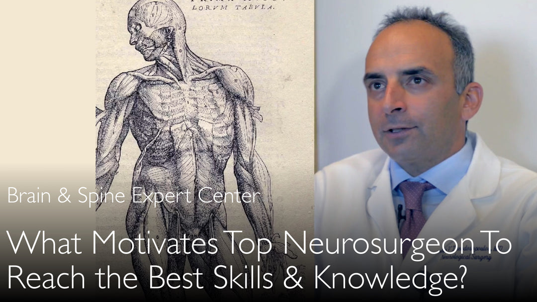
Dr. Philip Theodosopoulos, MD, asserts that a deep understanding of microsurgical anatomy is the fundamental building block for any neurosurgeon striving for world-level excellence. He traces the origins of modern surgery back to Renaissance pioneers like Andreas Vesalius, who emphasized the critical importance of human dissection. This historical perspective underpins his belief that surgeons must be grounded in anatomical knowledge above all else. Dr. Theodosopoulos argues that technical skill alone is insufficient without this foundational understanding.
Research Methods in Anatomical Study
Dr. Philip Theodosopoulos, MD, employs a multifaceted research approach to deconstruct the complexities of the skull base. His prolific work involves meticulous studies on dry human skulls using advanced 64-slice computed tomography (CT) imaging. For example, one study analyzed 84 dry skulls, while another conducted a morphometric analysis of 100 skulls using CT scans and Brain Lab navigation systems.
He complements this radiological analysis with hands-on endoscopic and microsurgical dissections. His research utilizes formalin-fixed cadaver heads and specimens to practice and refine surgical approaches, such as those through the middle turbinate, inferior turbinate, and maxillary sinus via the Caldwell-Luc approach.
Why Normal Anatomy is a Surgical Challenge
A central tenet of Dr. Theodosopoulos's philosophy is that normal anatomy itself is a primary challenge in surgery. He provides a stark warning: "Normal anatomy can reduce a great surgeon to a mediocre surgeon." During operations, surgeons most frequently encounter normal anatomical structures, not pathological ones. Without an exhaustive, intimate knowledge of every nuance and common variation, a surgeon's performance can be severely compromised.
This is why operating on patients alone is not sufficient for skill development. Dr. Philip Theodosopoulos, MD, explains to Dr. Anton Titov, MD, that a surgeon must go beyond the operating room to continuously study and relearn anatomical relationships to avoid being sub-optimal.
Cadaveric Dissection for Skill Development
Dr. Philip Theodosopoulos, MD, champions cadaveric dissection as an indispensable tool for honing surgical skills and innovating safely. He states that practicing new techniques in a simulated environment or on cadaveric material is a ethical imperative before attempting them on patients. Taking untested techniques straight to a patient amounts to "human experimentation," where the surgeon may not yet be proficient.
This method allows surgeons to gain extensive experience without risk to patients. It is the only way to safely "push the envelope" of what is surgically possible, understand the precise limits of different approaches, and fully comprehend the risks involved in a procedure.
Endoscopic Approaches and Anatomical Perspectives
A significant focus of Dr. Theodosopoulos's recent work is on mastering the anatomy for endoscopic endonasal cranial base surgery. He notes that familiar anatomy, like the skull base, looks entirely different when approached from below (endonasally) compared to a traditional cranial approach. While the anatomy itself does not change, the perspective and surgical corridors do.
This requires dedicated study to understand the limitations and relationships of structures from this specific vantage point. His research into areas like the internal carotid artery, optic strut, and inferior orbital fissure from an endoscopic view is crucial for developing safe and effective minimally invasive techniques.
The Imperative of Continuous Learning for Surgical Mastery
For Dr. Theodosopoulos, the pursuit of anatomical knowledge is not a phase of training but a lifelong commitment. He emphasizes that this rigorous, continuous study is crucial not only for training surgical residents and fellows but for "training ourselves." It is an ongoing process that every surgeon must engage in throughout their career.
This dedication to perpetual learning is what separates good surgeons from truly leading ones. Dr. Anton Titov, MD, highlights that this level of dedication is rare, comparing Dr. Theodosopoulos's work to following in the steps of Leonardo da Vinci. This relentless pursuit ensures that a surgeon remains grounded, understands their limits, and ultimately provides the highest standard of care to their patients.
Full Transcript
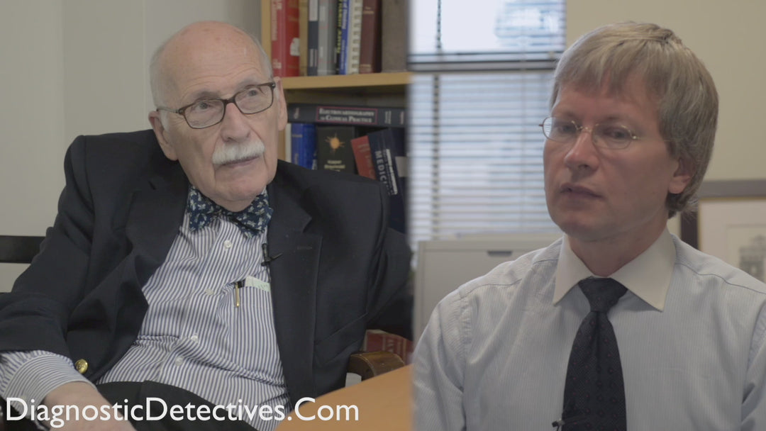
Dr. Anton Titov, MD: What motivates leading surgeons to excel in work and in life? A leading cerebrovascular and skull base neurosurgeon shares his passion and dedication.
Dr. Philip Theodosopoulos is following the path of Andreas Vesalius and Leonardo da Vinci. Professor Philip Theodosopoulos meticulously studies the surgical microanatomy of the skull base. He uses modern methods of CT scans to better understand every nuance of skull base anatomy.
"Surgeons mostly see normal anatomy when they operate on patients. Normal anatomy can reduce a great surgeon to a mediocre surgeon." "To be a superior surgeon, you have to go back to the basic building blocks of anatomy."
"In 84 dry human skulls, imaging studies were performed by 64-slice computed tomography." What motivates leading surgeons? Dr. Anton Titov, MD. What drives a highly skilled surgeon to continue studying the dissection anatomy of the skull? Dr. Philip Theodosopoulos, MD.
A video interview with a leading expert in skull base neurosurgery. Microsurgical anatomy of the skull is necessary to improve surgical skill. Surgeons mostly see normal anatomy when they operate on patients. Normal anatomy can reduce a great surgeon to a mediocre surgeon.
A medical second opinion from a leading surgeon can confirm a tumor diagnosis. A medical second opinion also helps to choose the best treatment for cancer or a benign brain tumor. Dr. Anton Titov, MD. Seek a medical second opinion on a brain tumor and be confident that your treatment happens according to leading international standards.
Dr. Philip Theodosopoulos, MD: Surgeons can only become better with more experience. But operating on patients is not sufficient. A surgeon has to continuously study dissection anatomy in his specialty. A neurosurgeon has to study brain anatomy on cadaveric heads.
To become a great surgeon, to push the envelope, a surgeon really has to go back to the basic building blocks of microsurgical dissection anatomy. What motivates leading surgeons? Becoming the best surgeon.
Dr. Anton Titov, MD: You have a particular interest in the delicate and very complex anatomy of the skull base. You prolifically publish this fascinating surgical anatomy research. Let me quote some titles from your six recent papers about the surgical anatomy of neurovascular skull base structures.
Endoscopic, endonasal variability in the anatomy of the internal carotid artery. Anatomic variation of the optic strut: classification schema, radiologic evaluation, surgical relevance. Dr. Anton Titov, MD. Anatomy of the inferior orbital fissure: implications for endoscopic cranial base surgery. Anatomic study of the prechiasmatic nucleus and its surgical implications.
Dr. Philip Theodosopoulos, MD: Anatomy of the optic canal: a computed tomography study of endoscopic nerve decompression. Endoscopic anatomy of the petrous segment of the internal carotid artery.
Let me also quote some methods that you use in some of these studies: "In 84 dry human skulls, imaging studies were performed by 64-slice computed tomography." Endoscopic endonasal dissections were performed in six formalin-fixed cadaver heads.
Morphometric analysis of 100 skulls was conducted using CT scans and the Brain Lab. Four patients underwent procedures that exposed the maxillary strut. In 10 cadaveric specimens, microanatomical and endoscopic dissections were performed.
Dr. Philip Theodosopoulos, MD: Dissections were done via approaches in the middle turbinate and the inferior turbinate. Dissections were performed also via the Caldwell-Luc treatment through the maxillary sinus.
These quotes from your work and your studies vividly demonstrate the degree of dedication. They show the need for continuous honing of surgical skills. They also show your understanding of very specific and delicate surgical anatomy.
Dr. Philip Theodosopoulos, MD: Such technical skills are required for a surgeon who wishes to have world-level excellence in his field. Not many surgeons today are literally following the steps of Leonardo da Vinci. Not many surgeons do their own anatomical research today.
Dr. Anton Titov, MD: What drives you in these rigorous pursuits? How does it help you in your clinical practice?
Dr. Philip Theodosopoulos, MD: It is interesting. Microsurgical anatomy has been my primary interest in research. My other interest is clinical outcomes research.
Medicine may have started with Hippocrates and Galen. But surgery really started with Andreas Vesalius and patients in the Middle Ages and Renaissance. In the Renaissance, they actually took an interest in dissecting the human body.
It is the only method how surgeons can really stay grounded in what is important in surgery. We need understanding that even the most experienced surgeons are reduced to mediocre surgeons so often by the normal anatomy that we encounter.
It goes without saying that for a surgeon to be good, a surgeon has to really study a lot.
Dr. Anton Titov, MD: The studying doesn't come only from the patients you see. Because there is just no method that you can see this many patients or do this many surgeries. To some degree, it ends up being human experimentation.
Sometimes you are not good at what you are devising. All of these new techniques that we have.
Dr. Philip Theodosopoulos, MD: Sometimes you take the new techniques straight to the patient instead of having done that many, many times in cadaveric material. Or you practiced new techniques in a simulated environment.
You are not able to say that we are safe to perform a new surgical technique on a patient. That is what we see in this anatomic work. We see that we can only become better surgeons by experience.
If you want to push the envelope, you really have to go back to basics of surgical methods. You have to go back to the basic building blocks of anatomy. You have to understand anatomy very well.
You have to understand the relationships between anatomical structures. The most recent work we have done is focusing on the endoscopic methods to treat the skull base.
Skull base that we have seen thousands of times from the leading. Skull base that we see from the subcranial area. It looks so different when you come from below. It is the same anatomy.
Anatomy doesn't change whether the method we come from below, from the leading, or from the side. You have to study and understand the limitations, relationships, et cetera, from the specific treatment that you are having.
Only then you are not going to be good. You are not going to understand the limits. You are not going to understand the risks you are taking. Then you are going to be sub-optimal in your surgical skills.
In training, it is crucial for all of us. It is not only crucial in training our surgical residents or fellows.
Dr. Anton Titov, MD: But in training ourselves, in training all of us, it is imperative that we have to do that. We have to study surgical anatomy continuously and rigorously.
What motivates leading surgeons? How to achieve leading surgical skills? A video interview with a leading expert in skull base neurosurgery. Surgical excellence and anatomy.